4° INCONTRO SULL’ECOSISTEMA TOSCANO PER L’INNOVAZIONE - SPOKE 1
Auditorium (Edificio A)
CNR - Area della Ricerca di Pisa
SPOKE 1: ADVANCED RADIOTHERAPIES AND DIAGNOSTIC IN ONCOLOGY
Le patologie oncologiche rappresentano una delle principali sfide per la sanità pubblica. L’innovazione scientifica e tecnologica è cruciale per affrontare questa emergenza, migliorando l’efficacia delle terapie e riducendo gli effetti collaterali. La radioterapia è un pilastro nel trattamento di molti tumori, coinvolgendo più della metà dei pazienti oncologici. Tuttavia, la ricerca attuale suggerisce la necessità di superare gli approcci tradizionali, esplorando nuove modalità di somministrazione delle radiazioni per ottimizzare i risultati clinici.
Tra le tecniche emergenti, l'effetto "FLASH" si distingue per la sua capacità di ridurre il danno ai tessuti sani attraverso la somministrazione ultra-rapida di alte dosi di radiazioni, garantendo però la stessa efficacia terapeutica contro le cellule tumorali rispetto alle modalità di somministrazione convenzionali. Questo rappresenta un passo avanti nelle terapie avanzate, aprendo la possibilità di trattare forme tumorali più resistenti e riducendo significativamente i tempi complessivi di trattamento, con benefici anche in termini di sostenibilità del sistema sanitario.
A distanza di un anno dal nostro ultimo incontro, il meeting 2024 dello Spoke 1 offre l’occasione di riflettere sui progressi ottenuti e di condividere i risultati delle attività di ricerca, che spaziano dall’utilizzo di tecnologie innovative in radioterapia all'applicazione di nuovi radiofarmaci e radiotraccianti per una diagnosi più precisa e mirata. Gli avanzamenti fatti nel campo delle terapie oncologiche hanno coinvolto un’ampia rete di competenze multidisciplinari, che includono fisici, biologi, ingegneri e clinici, tutti collaborando verso un comune obiettivo.
Il meeting sarà anche l'occasione per discutere le prospettive future e i nuovi progetti nell’ambito dello Spoke 1, il cui scopo è continuare a integrare le migliori innovazioni nel campo della radioterapia e della diagnostica oncologica. Lo Spoke 1, parte del più ampio progetto THE (Tuscany Health Ecosystem), mira a consolidare la Toscana come punto di riferimento per la ricerca e l’applicazione delle scienze della vita, promuovendo sinergie tra ricerca scientifica e applicazioni cliniche.
albert COMELLI
Alessandra FLORI
Alessandra RETICO
Alice USAI
Antonino FORMUSO
Barbara RICHICHI
Beatrice D'ORSI
Beatrice DI MARCO
Cinzia TALAMONTI
Claudia KUSMIC
Claudia MARTINI
Costanza FABBRI
Cristina SPALLETTI
Daniele PANETTA
David GREGOCKI
Debora PETRONI
Eduarda MOTA DA SILVA
Eleonora VANNINI
Elisabetta BIANCHINI
Elisabetta TOGNONI
Emma BUZZIGOLI
Enrica STRETTOI
Fabrizio CARTA
Felicia PELAGALLI
Francesca USALA
Francesco FAITA
Francesco MAZZINI
Gabriele SANSEVERO
Gian Michele RATTO
Gioele RENZI
Giorgio DE NUNZIO
Giulia FURINI
Guido GIORGI
Jessica DE GIOVANNI
Laura RESTANI
Leonida Antonio GIZZI
Lorenzo FULGENTINI
Luca LABATE
Luca MENICHETTI
Margherita MAFFEI
Maria Grazia ANDREASSI
Mariagrazia CELENTANO
Mario COSTA
MAURIZIO GIOVANNI PORTALURI
Melissa SANTI
Michela POLI
Milena RIZZO
Raffaella MERCATELLI
Riccardo CICCHI
Roberta MEZZENA
Silvia LANDI
Silvia SELLERI
Simon VLACHOS
Simona PICCININI
Simone CAPACCIOLI
Vinoshene PILLAI RAJAN
Viviana BENFANTE
- +38
-
-
09:00
→
10:00
SESSION 1 - SPOKE COORDINATION and MANAGEMENT Auditorium (Edificio A)
Auditorium (Edificio A)
CNR - Area della Ricerca di Pisa
Via G. Moruzzi, 1 Pisa-
09:00
Spoke 1 : Overview of activities and emerging plans for the future 20mSpeaker: Leonida Antonio GIZZI (Istituto Nazionale di Ottica, Pisa)
-
09:20
Breve aggiornamento su attività relative a regolatorio dei dispositivi medici - spoke 5 20m
The presentation will briefly provide an update about spoke 5 (IMPLEMENTING INNOVATION FOR HEALTHCARE AND WELL-BEING) activities with a focus on the medical device regulation tasks.
Speaker: Dr. Elisabetta BIANCHINI (IFC-CNR) -
09:40
La gestione del progetto: obiettivi e risultati raggiunti, azioni future 20mSpeaker: Donata FORNACIARI
-
09:00
-
10:00
→
10:30
Coffee break 30m Aula 29
Aula 29
CNR - Area della Ricerca di Pisa
-
10:30
→
11:15
SESSION 2 - SPOKE1: PROGRESS OF SUBPROJECTS "Clinical" Auditorium (Edificio A)
Auditorium (Edificio A)
CNR - Area della Ricerca di Pisa
Via G. Moruzzi, 1 Pisa-
10:30
Preliminary evaluation of Flash electron Radiotherapy in the treatment of Uveal melanoma 15m
Various in vivo studies proved that the FLASH treatment results in a decrease of radiation-induced normal tissue toxicity, compared to conventional treatment, while having the same biological effect on tumoral tissue.
The aim of this study is to verify the possibility of using FLASH electron Radiotherapy as an alternative treatment modality for localized uveal melanoma. This is a highly radioresistant tumor which constitutes the most common type of ocular disease in adults and is generally treated with organ-removal approaches (surgery) or organ-conservative techniques (such as Stereotactic RadioTherapy - SRT), depending on the overall tumor dimensions. This will be possible exploiting ElectronFlash (EF) LINAC situated in Santa Chiara Hospital, Pisa, which can deliver 7MeV-9MeV electrons beams to the target in both FLASH (>40Gy/s) and conventional (2Gy/min) regimes.
The main steps of the project are: i) validation of the EF simulation and optimization on patient CTs, ii) implementation of the FLASH sparing effect in simulations according to in vitro/vivo experimental results, iii) development of a robust method of comparison between FLASH and Stereotactic Radiotherapy (SRT) based on dosiomics and advanced statistics.
A dose deposition code based on EGSnrc MonteCarlo algorithm was developed to simulate EF. Some tests were conducted using flashDiamond detector, to verify the simulated dose distribution in water. Also, a dose distribution calculation on selected clinical cases was performed: patients with different tumor sizes and positions were considered, using different beam sizes and fields geometry to optimize the target coverage.
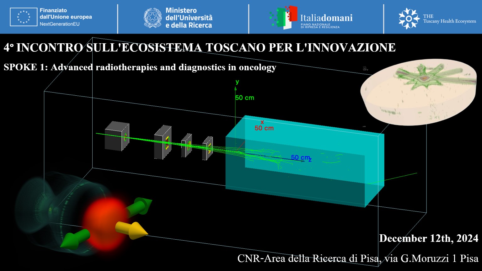
This analysis demonstrated a good agreement between the simulated and measured dose distributions. However, it soon became evident that these energies were insufficient to cover deep targets (>2cm along the beam axis). To overcome this limitation, virtual energy spectra were generated, starting from the real 9MeV spectrum and extending up to 30MeV. By employing these new energy spectra, it was possible to achieve better target coverage, using at least the 90% of the measured dose at the build-up point (Figure 1).
The simulated EF dose distributions will be further optimized. Simultaneously, radiobiological studies are being performed to quantify the sparing effects generated by FLASH-RT.
The final aim of this study is to evaluate the effectiveness of EF “virtual” treatment on selected patients and to make a direct quantitative comparison with SRT, with the help of dosiomics and advanced statistics.
This work was funded by Piano Nazionale di Ripresa e Resilienza (PNRR), Missione 4, Componente 2, Ecosistemi dell’Innovazione–Tuscany Health Ecosystem (THE), Spoke 1 “Advanced Radiotherapies and Diagnostics in Oncology”—CUP I53C22000780001.
We also thank Fondazione Pisa for funding CPFR with the grant “prog. n. 134/2021”.Speaker: Mariagrazia CELENTANO (INFN, Pisa Section, Pisa, Italy) -
10:45
Radiofarmaci per uso clinico prodotti nell'officina farmaceutica: stato di avanzamento 15m
L’obiettivo generale del WP8 è quello di fornire radiofarmaci innovativi PET, per uso clinico, da utilizzare nella diagnosi e nel follow-up dei gliomi e di altre patologie oncologiche.
L’officina farmaceutica produce quotidianamente i due radiofarmaci (18F-FDG and 18F-FColina) previsti nel milestone 2 con le seguenti Marketing Authorizations:
• Fluorocolina (18F) Curium Italy 225 MBq/mL soluzione iniettabile (AIC: 045030)
• FLUODEOSSIGLUCOSIO (18F)-IBA 185 MBq/mL soluzione iniettabile (AIC: 036946).- Come primo risultato del progetto PNRR del WP8, si è concluso il trasferimento tecnologico della produzione di FLUODEOSSIGLUCOSIO (18F)-IBA 185 MBq/mL sulla piattaforma Trasis mentre per 18F-Fluorocolina è stato avviato il trasferimento tecnologico sulla piattaforma Trasis che si concluderà nel Q1 del 2025.
- Trasferimento tecnologico e autorizzazione AIFA del radiofarmaco 18F-Fluoroetiltirosina (18F-FET o IASOGLIO): il risultato principale ottenuto dal WP8 è stato Il trasferimento tecnologico di IASOGLIO®, completato a febbraio 2024, e il rilascio da parte di AIFA dell’autorizzazione alla produzione secondo GMP di tale radiofarmaco. Ad oggi, la 18F-FET non era disponibile in Italia per la grande distribuzione e veniva importata dall'estero. L’officina farmaceutica IFC-CNR è il primo e unico sito produttivo italiano autorizzato da AIFA a produrre 18F-FET ed è in corso presso EMA la procedura di mutuo riconoscimento per avviare la grande distribuzione. Il progetto “PROFETI”, proposto da Curium Pharma nell'ambito dei bandi a cascata THE, è stato finanziato e consentirà l'estensione del trasferimento tecnologico di IASOGLIO® ai siti produttivi di Roma Tor Vergata, Milano Istituto Europeo di Oncologia e Udine.
- Produzione del radiofarmaco sperimentale 18F-FAPI74. Il radiofarmaco sperimentale 18F-FAPI74 è stato sintetizzato grazie all’accordo stipulato con l’azienda statunitense SOFIE, detentrice del brevetto del precursore per la sintesi di 18F-FAPI74. SOFIE collabora con numerose istituzioni accademiche a livello globale per dimostrare la potenziale utilità clinica di FAPI tramite il FAPI GLOBAL OUTREACH PROGRAM. Per entrare in questo importante programma, insieme al nostro partner di progetto la Medicina Nucleare di AOUPI, abbiamo inviato a SOFIE due protocolli clinici per la loro approvazione. Due protocolli clinici, uno per lo studio del glioma (TARG-Glioma) e uno per lo studio dell'epatocarcinoma (DETECT) sono stati approvati da SOFIE. Il primo lotto di 18F-FAPI74 è stato sintetizzato, stiamo preparando l’IMPD e in questo momento è in corso la redazione della documentazione per procedere alla richiesta di autorizzazione ai trials clinici attraverso la piattaforma CTIS.
- Trasferimento Tecnologico 18Fluoro-DOPA. Il trasferimento tecnologico della Fluoro-DOPA è stato completato (Settembre 2024), tutti i metodi analitici sono stati validati e la richiesta di autorizzazione ad Autorizzazione ad AIFA per la produzione su larga scala è in corso
Speaker: Letizia GUIDUCCI -
11:00
Towards the clinical applications of Flash Radiotherapy. An overview of the 1.5.4/1.5.8 deliverables 15m
In this presentation, the deliverables 1.5.4, 1.5.5, 1.5.6, 1.5.7 and 1.5.8 will be discussed.
The ElectronFlash linac performances have been characterized using both active and passive dosimeters and the reference conditions have been established.
We developed and validated, trough experimental data, a framework based on MonteCarlo simulations tuned with the ElectronFlash main parameters. Our simulations are performed on CT scans of both animals and patients. Regarding animals, even though the simulations led us to dose maps to better plan the experimental set-up, a custom imaging and positioning system is necessary and will be developed through cascade tenders.
To achieve the construction and use of a positioning and centering system for patients, it is necessary to first go through a system suitable for small animals. We also developed a treatment planning system based on MonteCarlo simulations validated using a water phantom. We can vary among all ElectronFlash parameters. Additionally, we are developing a beam shaper device to further optimize the PTV coverage without affecting the beam dosimetric parameters. In the optimization phase, we can also account for water-equivalent bolus. Additionally, the TPS can calculate the key beam parameters to use in FLASH irradiations, such as the average dose-rate, instantaneous dose-rate and dose-per-pulse along with dose volume histograms (DVHs) to evaluate PTV and OARs coverage. Once obtained the experimental thresholds (functions) that describe the FLASH sparing effect, we can incorporate them into the evaluation of DVHs to obtain the RBE-weighted DVHs.Speaker: Andrea CAVALIERI (The Center for Instrument Sharing of the University of Pisa (CISUP))
-
10:30
-
11:15
→
13:30
SESSION 3 - SPOKE1: PROGRESS OF SUBPROJECTS "Technology and Simulations" Auditorium (Edificio A)
Auditorium (Edificio A)
CNR - Area della Ricerca di Pisa
Via G. Moruzzi, 1 Pisa-
11:15
Update on requirements collection and first prototypes of the data platform 15m
In questa presentazione verrò fornito un aggiornamento sullo sviluppo della piattaforma per lo storage e l’analisi dei dati nel progetto THE. Verrà introdotto un prototipo iniziale basato su XNAT, che abbiamo sviluppato sfruttando l’infrastruttura dell’INFN e collaborando anche con lo Spoke 4, per gestire immagini e dati di microscopia. Verrà mostrata l’interfaccia relativa a questo prototipo anche a scopo dimostrativo, per illustrare le funzionalità della piattaforma. Si discuterà il lavoro di raccolta dei requisiti dai vari gruppi che stiamo portando avanti, con la loro traduzione in uno schema organizzativo dei dati implementabile in XNAT. Inoltre, verrà fornita una panoramica delle potenziali analisi basate su tecniche di Intelligenza Artificiale, che potranno essere utilizzate per assistere l’analisi e l’interpretazione dei dati raccolti.
L'obiettivo è creare una piattaforma scalabile e integrata per supportare le attività di ricerca del progetto, in maniera in realtà trasversale a diversi sottoprogetti.Speaker: Camilla SCAPICCHIO (INFN sezione di Pisa) -
11:30
A minimalistic phenomenological model of the FLASH effect. 15m
In phenomenological stochastic models of the biological cellular effect of ionizing radiation, e.g. the multiple target multiple hit model (MTMH), one uses the concept of threshold - the effect happens if at least M critical cellular targets are hit by at least N ionizing particles each, the effect doesn’t happen otherwise. In these models, a notable sigmoidal shape of the biological effect versus absorbed dose curve originates from the spatial inhomogeneity of the cellular regions hit by the radiation - some regions are hit many times and contribute to the possible cellular effect, the others do not. The active radicals and ions created by the ionizing particles and eventually (directly or indirectly) causing the biological damage also have spatially inhomogeneous concentration or, in other words, mesoscopic spatial fluctuations around the average value. The fluctuations of the radial concentration dissipate due to the diffusion and the recombination processes in competition with creation of new radicals due to dose deposition at a given dose rate, and this kinetics affects the eventual biological effect.
We present a simple theory of the diffusive relaxation of mesoscopic, non-thermodynamic fluctuations of radical concentration and its application to the dose rate (FLASH) effect in radiotherapy. The conclusions of the model are in qualitative agreement with the recent review [1] of the available experimental results of the FLASH sparring effect for normal tissue complications probability. In particular the model predicts the increase of the FLASH effect at higher doses and also the threshold (minimal dose) below which the FLASH sparring is not observed.[1] Böhlen TT, Germond JF, Bourhis J, Vozenin MC, Ozsahin EM, Bochud F, Bailat C, Moeckli R. Normal Tissue Sparing by FLASH as a Function of Single-Fraction Dose: A Quantitative Analysis. Int J Radiat Oncol Biol Phys. 2022 Dec 1;114(5):1032-1044. doi: 10.1016/j.ijrobp.2022.05.038. Epub 2022 Jul 8. PMID: 35810988.
Speakers: Igor BODRENKO (CNR-NANO) , Valentina TOZZINI -
11:45
The future of VHEE medical applications: what simulations are telling us 15mSpeaker: Costanza PANAINO (CNR INO)
-
12:00
VHEE generation and transport: experimental work 15m
In the last decades, Laser plasma acceleration (LPA) has seen a growing interest both for pure research purposes but also for being employed in applications. One of the key features is the possibility to reach high acceleration gradients, resulting in compact accelerators and reduced costs. Laser Wake-Field Acceleration (LWFA) is routinely used to produce energetic electrons and, by properly tuning the injection mechanism, high quality beams can be produced. At the Intense Laser Irradiation Laboratory (ILIL) a Titanium-Sapphire laser (6 J, 30 fs, 0.8 μm) is focused on a supersonic flow of Helium gas doped with Nitrogen at 1% so to trigger the production of Very High Energy Electron - VHEE (50-250 MeV) via the ionization injection mechanism granting high collimation and pointing stability. The latter can be further improved by means of magnetic beamline (MBL). We designed a modular and cost-effective MBL that thanks to its modularity can be adjusted to fit different purposes and beam energies.
The generated electrons are then let in air where they are characterized in terms of energy, dose and charge. To this purpose the diagnostic bench has been equipped with a magnetic spectrometer, an Integrating Current Transformer (ICT) and a set of radiochromic films holders both for studying the longitudinal dose deposition profile and have an estimate of the beam dose per each pulse. Once the electron beam has been fully characterized it can be used to irradiate biological samples.Speaker: Martina SALVADORI (CNR-INO) -
12:15
Spectral features of VHEE beams 15m
Accurately measuring the energy of very high-energy electron (VHEE) beams is crucial if they are to be considered for applications. Laser-driven electrons are notoriously known for beam pointing and divergence variations from shot-to-shot. We have developed a spectrometer system that accurately measures the energy, by simultaneously correcting for pointing and charge. We conducted laser wakefield acceleration experiments resulting in electrons reaching a maximum energy at above 400 MeV, with peak at ~165 MeV peak and ~65% energy spread. Such spectra, although broad, can prove sufficient for Flash radiotherapy if energies below 100 MeV are filtered out.
Speaker: Simon VLACHOS (CNR-INO) -
12:30
Correlation Study of Integrating Current Transformer Charge and Lanex Screen Emission Signal in High-Energy Beam Measurements 15m
Over the past ten years, laser plasma accelerators (LPA) have shown remarkable progress, influenced in part by advances in laser technology. Their ability to produce quasimonoenergetic electron beams with energies ranging from tens of MeV to tens of GeV in just a few millimeters to centimeters brought LPA to the attention of many scientific and industry fields. Arguably the most important of them is medicine, in a newly developed, so-called FLASH radiotherapy. In this regard, one of the important requirements that an electron beam generated via LPA has to fulfill is shot-to-shot stability in terms of energy, pointing stability, and charge. For this reason, experimental studies have been conducted at the ILIL, INO-CNR, in which electron beam charge has been directly measured by the integrating current transformer (ICT). The results from the measurements have been put into a correlation with the Lanex screen emission signal to obtain a calibration curve.
Speaker: David GREGOCKI (Consiglio Nazionale delle Ricerche - Istituto Nazionale di Ottica) -
12:45
Tomographic Reconstruction of Dose Distribution for 3D-Dosimetry in FLASH-RT using a Monolithic Plastic Scintillator Block 15m
This contribution presents the development of a 3D online dosimeter based on a plastic scintillating monolithic block, imaged with cameras to reconstruct the three-dimensional dose distribution using a tomographic algorithm. The study primarily relies on simulations to investigate the feasibility and performance of the proposed system.
The simulated detector consists of a 10 cm polyvinyl-toluene-based plastic scintillator block (EJ200, Eljen Technology, Texas) positioned along the beamline to absorb a 9-MeV electron beam. Scintillation light emitted from the detector is captured by three cameras, each coupled to a focusing objective, and positioned along three orthogonal axes to collect the light emitted along directions parallel and orthogonal to the beam axis. A maximum likelihood approach is employed in the reconstruction algorithm to derive 3D dose maps from lateral light projections.
In this contribution, we will present the performance of the detector and the algorithm, validated through simulations of the ElectronFlash LINAC accelerator at the Centro Pisano Flash Radiotherapy (CPFR) in Pisa. Additionally, the potential use of Cerenkov light emitted within a plastic cube as a signal for reconstruction will be briefly discussed.
Speaker: Eleonora RAVERA (UniPi) -
13:00
VHEE beam at sample and dosimetry: experimental approach (Milestone 1.1 and 1.6) 15m
This study presents the experimental advancements in the generation and dosimetric characterization of laser-plasma accelerated Very High Energy Electron beams (100–250 MeV) for radiobiological applications.
These beams, known for their deep tissue penetration and reduced sensitivity to anatomical inhomogeneities, have been studied to evaluate their potential in advanced radiotherapy applications.
The experimental work involved dosimetric measurements using stacks of EBT3 radiochromic films to map the dose distribution of a collimated electron beam with a 7 mm diameter. Dose delivery was evaluated for multiple ultra-high dose rate pulses. The characterization was performed to optimize irradiation and dose measurement for radiobiological samples.
3D-printed sample holders designed and produced for these experiments are described. These holders are adaptable to various containers (e.g., 0.5 ml Eppendorf tubes, 2 ml cryotubes, and 25 mm Petri dishes) and can integrate radiochromic dosimeters and stainless steel collimators to enhance irradiation precision. A motorized centering device enables precise sample positioning within the irradiation field. Additionally, a new holder with orthogonal angular movements and laser-guided alignment significantly improve beam alignment and irradiation homogeneity.Speaker: Simona PICCININI (ILIL CNR-INO) -
13:15
First radiobiology assays with UHDR VHEE beam 15m
This research investigates the radiobiological response and dose-dependent chromosomal damage in human lymphocytes exposed to very high-energy electron (VHEE) beams, employing the cytokinesis-block micronucleus (CBMN) assay—an internationally recognized biodosimetric method endorsed by the International Atomic Energy Agency (IAEA). Micronuclei (MN) are small, extranuclear bodies that form when whole chromosomes or chromosomal fragments are not incorporated during mitosis. The presence of radiation-induced MN, typically assessed in blood lymphocytes, serves as an indicator of unrepaired or misrepaired DNA double-strand breaks. Human peripheral blood is an ideal non-invasive biological sample for cytogenetic tests aimed at detecting DNA damage following irradiation. T-lymphocytes, known for their heightened sensitivity to radiation, are particularly beneficial for studying induced chromosomal damage. These cells are irradiated while synchronized in the G0 phase of the cell cycle, a resting state prior to replication. Following stimulation with phytohemagglutinin (PHA), a mitogenic agent, T-lymphocytes enter mitosis, enabling us to assess the effects of radiation on these cells.
Furthermore, we investigated the impact of VHEE on telomere length and mitochondrial DNA. Telomeres are considered "hallmarks of radiosensitivity" and are primary targets for reactive oxygen species (ROS), which contribute to their progressive shortening and subsequent genomic instability. Mitochondrial DNA (mtDNA) is also vulnerable to ionizing radiation and is more susceptible to damage than nuclear DNA due to the absence of protective histones and limited DNA repair capacity.
Our preliminary findings indicate a radiation dose-response relationship in chromosomal damage, evidenced by an increase in the frequency of MN with higher radiation doses. This phenomenon applies to both targeted effects, where damage occurs in irradiated cells, and indirect effects, characterized by biological changes in non-irradiated cells as a result of signals from irradiated cells. Additionally, we observed radiation-induced telomere shortening due to VHEE exposure; however, no significant difference was found in the analysis of mtDNA copy number (mtDNAcn), a surrogate marker of mitochondrial dysfunction.Speaker: Andrea BORGHINI (IFC CNR)
-
11:15
-
13:30
→
15:00
Lunch (Buffet) 1h 30m Aula 29
Aula 29
CNR - Area della Ricerca di Pisa
-
15:00
→
16:15
SESSION 4 - SPOKE1: PROGRESS OF SUBPROJECTS "Pre-Clinical" Auditorium (Edificio A)
Auditorium (Edificio A)
CNR - Area della Ricerca di Pisa
Via G. Moruzzi, 1 Pisa-
15:00
An in vitro platform to investigate the FLASH effects on melanoma and normal skin cells 15m
Background and Aims
Melanoma is the most lethal type of skin cancer due to its aggressive nature. Surgery and immunotherapy are the mainstay of treatment, but radiotherapy (RT) can also be a useful treatment option. FLASH-RT, which reduces radiation-induced side effects while maintaining the same level of tumor control as conventional RT, could present a paradigm shift, allowing to increase the total dose and the dose per fraction. Our aim is to test an in-vitro platform of increasing complexity of melanoma and normal cells to investigate the effects of FLASH-RT.Methods
Normal (SGBS, HaCat, NHDF) and cancer cell lines (A375) were cultured in 2D and through a two-layered-3D-bioprinted model. A dose range of 4-16 Gy was delivered in both CONV and FLASH (240 Gy/s) mode using the "Electron-Flash", a low energy electron LINAC with a triode-gun. Biological parameters were monitored (DNA damage, membrane permeability, colony growth pattern, cell death, metabolic and energetic profile) to obtain a comparative evaluation of radiobiological effects resulting from equal doses of CONV and FLASH on both normal and cancer cells.Results
At all doses tested FLASH and CONV-RT show equal efficacy in reducing melanoma cells survival. Normal cells prove to be radioresistant with preserved/slightly reduced survival in all conditions and a moderate FLASH effect revealed by a lower mortality at the dose of 8 Gy. SGBS adipogenesis, used as a read-out of function, was reduced by both types of RT, with CONV having a more pronounced impact.Conclusions
Our work achieves a cross fertilization effort between the experimentation of new technologies and the implementation of the state-of-the art of FLASH-RT, demonstrating a profound difference in the response of normal and cancer cells to both CONV and FLASH-RT. Our results demonstrate that the administration of FLASH-RT would lead to therapeutic effects on the tumor but limits the damage to healthy tissues.Speaker: Alice USAI -
15:15
Effects of Conventional and FLASH radiotherapy on glioblastoma and melanoma cells 15m
Radiotherapy is one of the most effective anti-tumor therapies, used in more than 60% of cancer patients at some point in their oncological treatment to eliminate/reduce the size of the tumor. At present, conventional radiotherapy (CONV-RT), the main approach used in clinic, presents some limitations, including the dose fractionation into several daily sessions and the risks for the surrounding normal tissue. Recently, an ultra-high-dose rate electron irradiation method termed FLASH radiotherapy (FLASH-RT) selectively spares healthy tissue while leaving unchanged the therapeutic effect on tumor cells. The radiobiological mechanism of FLASH-RT responsible for the FLASH effect have yet to be fully explored. Recent studies have shown that FLASH-RT can induce the protection of mammalian cells through transient hypoxia, potentially reducing reactive oxygen species (ROS) formation and DNA damage, thus decreasing toxicity to healthy tissues. Moreover, a deeper understanding of how calcium signaling is remodeled in some cancers and the consequences of calcium signaling on key events, such as proliferation, invasion and sensitivity to cell death, is needed. In fact, mitochondrial Ca2+ controls ATP synthesis, apoptosis, ROS generation and biosynthesis, and can determine the fate of the cell.
We aimed to examine the FLASH-RT-induced radiobiological damage occurring during and post irradiation, to understand the key processes involved and FLASH potential in therapy. Using combined population-based and single-cell imaging approaches, we investigated Ca2+ homeostasis, ROS formation, cell death, mitochondrial and cellular markers, gene expression and cancer metabolic reprogramming in murine glioma GL261 and melanoma B16-F10 cells compared to "non-tumoral control" cells using three increasing total irradiation doses (4, 8, 16 Gy).
Interestingly, our data demonstrated that both FLASH and CONV mode produced a significant raise in cellular ROS compared to the control cells in a dose dependent manner. At any dose and in all cells, CONV induced higher ROS production than FLASH treatment. Moreover, both FLASH and CONV resulted in a significant increase in intracellular Ca2+ compared to the control cells in different ways, at lower doses (4 and 8 Gy) for GL-261 cells, and only at 8 Gy for B16-F10 cells. Moreover, cell injury, significantly increased 72 h following both CONV- and FLASH-RT in a dose-dependent manner, indicating that both treatments induce damage to GL-261 and B16-F10 cells. However, FLASH-RT triggered more cell death at 4 Gy compared to CONV-RT. We next investigated the temporal (0 to 72 h post-irradiation) transcriptional activation of several genes involved in cell death, cell cycle arrest, senescence and autophagy observing that FLASH-RT induces greater transcriptional activation than CONV-RT 24h post-irradiation. Finally, different mitochondrial and cellular markers, and cancer metabolism were assessed using Flow Cytometry and Seahorse analysis highlighting the differences between FLASH and CONV mode and between non-tumoral control and cancer cells.Speaker: Beatrice D'ORSI (CNR-IN) -
15:30
Saving Neurons: The Sparing Effect of FLASH Radiotherapy 15m
Radiotherapy (RT) is a widely used treatment for various cancers. While it effectively targets cancerous cells, RT also damages the surrounding healthy tissue, particularly in sensitive regions like the central nervous system. This collateral damage poses significant challenges in maintaining neurological function.
Ultra-high dose-rate FLASH radiotherapy (FLASH-RT) has emerged as a promising advancement in oncology, demonstrating the ability to reduce normal tissue toxicity while preserving its potent anti-tumor efficacy—an effect known as the "FLASH effect."
In this study, we explore the neuroprotective sparing effect of FLASH-RT compared to conventional radiotherapy (CONV-RT) in healthy nervous tissue. Using animal models, we assess cognitive outcomes and the functional impact of these two radiation modalities.
Methodology:
• Radiotherapy Treatment: Whole-brain irradiation with a single dose (15 or 20 Gy) using an electron beam at either a FLASH dose rate of 257 ± 2 Gy/s or a CONV dose rate of 4 ± 0.02 Gy/s.
• Behavioral Assessments: Object Recognition Test (ORT), Y-Maze, and Visual Cliff tests.
We examined various neural regions, spanning sensory, motor, and cognitive structures. By longitudinally monitoring the animals, we found that FLASH-RT had a milder impact on healthy nervous tissue compared to CONV-RT.. Additionally, mice treated with FLASH-RT demonstrated a faster recovery, with behavioral and physiological parameters returning more rapidly to levels comparable to untreated/control mice.
These findings highlight the potential of FLASH-RT as a revolutionary approach in radiotherapy, minimizing neurological side effects while maintaining therapeutic effectiveness.Speaker: Gabriele SANSEVERO (IN CNR) -
15:45
FLASH radiation effects on ocular tissues 15m
Uveal melanoma is the most common primary intraocular malignancy in adults. Eye enucleation has long been the gold standard for treatment but adverse effects have shifted recommendations toward eye preservation and radiotherapy. This can also lead to complications like radiation retinopathy, retinal detachment and optic neuropathy. To overcome the limitations of conventional radiotherapy (CONVRT), flash radiotherapy (FLASHRT) might represent a promising alternative. This novel technology involves the ultrafast delivery of radiation at dose rates much higher than those used in CONVRT (40 Gy/s versus 0.5–5 Gy/min, respectively), causing less damage to healthy tissues but achieving similar disease control (the so-called flash effect).
In this study, we employed a dedicated Linear Accelerator (Linac) with a triode gun, enabling in vitro and in vivo studies and able to switch rapidly between ultra-high and conventional modalities under controlled conditions. We compared the effects of FLASH and conventional radiations in healthy ARPE-19 cells, modeling the human retinal pigment epithelium (RPE), and in the retina and RPE of living, healthy mice, using various radiation protocols. Our long-term aim is to develop a protocol for ocular melanoma treatment. We demonstrated the successful occurrence of a flash effect on ARPE-19 cells under specific doses and discovered novel effects of the radiations with the protocols used. Stemming from these data, we initiated an in vivo toxicity study on whole-brain irradiated mice using 20 Gy and 15 Gy with either conventional or flash methods, performing chronic and acute observations. Notably, we observed no significant differences in the morphology of the RPE from FLASH versus CONV mice in both acute and chronic groups. This may be due to a dose exceeding the range necessary to generate a flash effect; the outcomes of lower doses are presently under investigation. A morphological analysis of the retinal tissues labelled with antibodies against Iba1, a protein specifically expressed by microglia and macrophages, demonstrated a lower inflammatory response in the retina of animals treated with Flash compared to mice irradiated using conventional methods. Molecular analyses to confirm these data are currently in progress.Speaker: Beatrice DI MARCO (Istituto di neuroscienze, CNR Pisa) -
16:00
Radiotherapy-induced ultrastructural modifications of corneal collagen probed by SHG microscopy 15m
Second-harmonic generation (SHG) microscopy is based on a coherent non-linear scattering process that allows high-resolution deep-tissue imaging of biological structures having high hyperpolarizability and structural anisotropy. For this reason, SHG represents a powerful tool for imaging collagen and probing its hierarchical organization from molecular scale up to tissue architectural level. In this study, SHG microscopy is used to characterize collagen ultrastructural modifications in murine corneal buttons treated with conventional and FLASH radiotherapy (RT). This evaluation serves as a benchmark of the structural integrity of the cornea, which is essential for the initial passage of light into the visual system. Stacks of optical sections covering the full corneal thickness were characterized using image analysis methods and parameters describing the organization of collagen structures at a supramolecular level. The preliminary results using Electron Flash show an increased off-plane contribution of SHG emitters after RT, with a lower variation in FLASH-RT than in conventional-RT, when compared to control samples indicating potential tissue-sparing effects. Despite additional samples and a larger statistic would be required to confirm the results and to understand the potential of this methodology for monitoring dose-dependent modifications of collagen, this approach could become a useful tool for monitoring the effect of RT on connective tissues and for evaluating the impact of different ionization sources.
Speaker: Riccardo CICCHI (Istituto Nazionale di Ottica (CNR-INO))
-
15:00
-
16:15
→
16:45
Coffee break 30m Aula 29
Aula 29
CNR - Area della Ricerca di Pisa
-
16:45
→
17:45
SESSION 4 - SPOKE1: PROGRESS OF SUBPROJECTS "Pre-Clinical" Auditorium (Edificio A)
Auditorium (Edificio A)
CNR - Area della Ricerca di Pisa
Via G. Moruzzi, 1 Pisa-
16:45
Evaluating FLASH and Conventional radiation response on brain-resident microglia 15m
Ultra-high dose-rate FLASH radiotherapy (FLASH-RT) has emerged as a promising approach in oncology, potentially enhancing the therapeutic index by minimizing normal tissue toxicity while maintaining anticancer efficacy compared to conventional radiotherapy (CONV-RT). However, the radiobiological effects on cranial radiotherapy are yet to be fully understood. This study investigates the in vivo effects of FLASH-RT and CONV-RT on brain-resident microglia, which play a crucial role in maintaining central nervous system (CNS) homeostasis and serve as key markers of neuroinflammation. To study microglial response towards irradiation, we employed intravital two photon imaging in mice expressing green fluorescent protein (GFP) under the fractalkine receptor promoter (CX3CR1GFP/+), following cranial window implantation. Two weeks post-implantation, the animals received a single cranial irradiation dose of 15 Gy using electron beams at either a FLASH dose rate of 257 ± 2 Gy/s or a CONV dose rate of 4 ± 0.02 Gy/s. In vivo two-photon imaging was conducted at multiple time points: pre-irradiation, 1–2 hours post-irradiation, and on days 7, 14, 21, and 28 post irradiation. Our results reveal that both FLASH-RT and CONV-RT induced notable changes in microglial dynamics and morphology, with CONV-RT exerting a more pronounced impact. These findings provide essential insights into the effects of radiotherapy on brain-resident microglia, contributing to our understanding of CNS responses to radiation and paving the way for improved radiotherapeutic approaches.
Speaker: Vinoshene PILLAI RAJAN (Institute of Neuroscience, CNR Pisa) -
17:00
Comparative analysis of FLASH and conventional radiotherapy in a murine model of melanoma and healthy animals 15m
Aim
We aimed to assess and compare the effects of FLASH-RT and CONV-RT on tumor growth in a melanoma model and on skin toxicity and systemic responses in healthy mice.Methods
A melanoma model was established by injecting B16F10 cells intradermally into the left leg of mice. After five days, mice were irradiated with either 19 Gy or 35 Gy using FLASH (940 Gy/s) or CONV modes with an Electron-Flash LINAC. Tumor volume, vital parameters, energy balance, metabolic and inflammatory markers were monitored. In a parallel group of healthy mice, the same doses and modalities were used to irradiate the left leg. Following irradiation, animals were monitored over time (5 wks), for skin toxicity and systemic effects.Results
Tumor Control: Both FLASH-RT and CONV-RT effectively controlled tumor growth, with higher efficacy at the higher radiation dose (35Gy). At 35 Gy, 25% of mice presented complete tumor remission regardless of the modality. Survival rates were significantly improved in irradiated mice compared to non-irradiated controls (P<0.0001), with the group of animals irradiated at 35Gy showing an higher survival rate compared to those irradiated with 19Gy. Although both modalities present similar efficacy, FLASH -RT group shows less damage to the skin surrounding tumor.Skin Toxicity and systemic effects on healthy mice: Healthy mice irradiated with 19 Gy experienced reduced food intake compared to control mice, with a more pronounced difference in the CONV-RT group. Alopecia and fur discoloration were also observed at an earlier time point in CONV-RT compared to the FLASH-RT group.
At 35 Gy, irradiated animals showed a lower weight gain compared to control animals, but differences between FLASH-RT and CONV-RT in food intake or body weight were not significant. Skin damage was more severe in the CONV-RT group, necessitating earlier euthanasia. CONV-RT mice had elevated IL-6 and ghrelin levels and reduced leptin and insulin levels compared to controls and FLASH RT miceConclusion
FLASH-RT demonstrated equivalent tumor control compared to CONV-RT with a notable sparing effect on normal tissue. The systemic markers profile indicates that CONV-RT may induce a starvation state of perceived starvation, potentially compromising long-term energy homeostasis. FLASH-RT's tissue-sparing properties merit further investigation for clinical translation.Speaker: Eduarda MOTADASILVA -
17:15
Development of multimeric radiotracers for the design of tumor microenvironment-selective theranostic agents for cancer diagnosis and therapy 15m
Cancer theragnostics is an emerging field of nuclear medicine that allows the integration of imaging and radiotherapy. It is based on the use of a pair of radiopharmaceuticals with the same molecular structure, one containing a positron-emitting isotope (such as fluorine-18 (18F) or gallium-68 (68Ga)) for PET imaging, aiding disease staging, therapy selection and monitoring. The other containing an alpha- or beta-emitting nuclide, such as lutetium-177 (177Lu), for targeted radiotherapy [1]. In recent years, several selective radiotracers have been developed and approved for clinical use based on differences in the overexpression of proteins on the cell surfaces of normal and malignant cells [2]. However, standard monospecific radiotracers are often ineffective due to the heterogeneity of the tumour and the complex interactions between the neoplastic cells and the surrounding microenvironment [3]. These radiotracers also suffer from limited tumor retention, reducing their therapeutic utility [4]. To address these limitations, multimeric agents able to hit multiple targets simultaneously have been developed, enhancing tumor specificity, uptake, sensitivity, and retention compared to traditional agents [5] – [7].
This project aims to develop novel classes of multimeric theragnostic agents selectively targeting the tumor microenvironment, utilizing integrin ligands (RGD peptides), fibroblast activation protein inhibitors (FAPI), and carbonic anhydrase ligands (CA IX/XII). These compounds will be labelled with 18F for in vivo PET imaging to assess tumor uptake and biodistribution and at the University Medical Center Groningen (UMCG) will be labelled with 177Lu for in vitro studies to assess cellular uptake, affinity, specificity and antitumour activity. For both in vivo imaging studies and in vitro therapeutic effect assessment, the performance of multimeric compounds will be compared to their monomeric counterparts.
Currently, radiolabelling protocols with 18F have been optimised for monomeric radiotracers. In detail, decay-corrected radiochemical yields of 30.9 ± 2.5 % and 28.3 ± 3 0 %, with a molar activity of 3.0 ± 0.2 GBq/umol and 1.9 ± 0.7 GBq/umol, were obtained for [18F]AlF-NOTA-FAPI-04 and [18F]AlF-NOTA-RGDfK, respectively. Both radiotracers showed a radiochemical purity (RCP) ≥97 % and stability in PBS and FBS for two hours. For [18F]AlF-NOTA-CA IX/XII, although optimal radiolabelling conditions were identified, the purification failed to meet the standards for animal use (RCP >95%), requiring further purification of the precursor by the NERUOFARBA department of the University of Florence. In addition, the first in vivo PET imaging studies were carried out on a B16F10 syngeneic mouse model of melanoma with the monomeric [18F]AlF-NOTA-FAPI-04 and [18F]AlF-NOTA-RGDfK radiotracers with the aim of highlighting their tumour uptake and biodistribution for a subsequent comparative study with the corresponding multimeric radiotracer."[1] Zoi V, Giannakopoulou M, Alexiou GA, Bouziotis P, Thalasselis S, Tzakos AG, Fotopoulos A, Papadopoulos AN, Kyritsis AP, Sioka C. Nuclear Medicine and Cancer Theragnostics: Basic Concepts. Diagnostics (Basel). 2023 Sep 26;13(19):3064. doi: 10.3390/diagnostics13193064. PMID: 37835806; PMCID: PMC10572920.
[2] Y.-Y. Huang, "An Overview of PET Radiopharmaceuticals in Clinical Use: Regulatory, Quality and Pharmacopeia Monographs of the United States and Europe," in Nuclear Medicine Physics, IntechOpen, 2019. doi: 10.5772/intechopen.79227.
[3] D. F. Quail and J. A. Joyce, "Microenvironmental regulation of tumor progression and metastasis," Nat. Med., vol. 19, no. 11, pp. 1423– 1437, Nov. 2013, doi: 10.1038/nm.3394
[4] C. D. van der Heide and S. U. Dalm, “Radionuclide imaging and therapy directed towards the tumor microenvironment: a multi-cancer approach for personalized medicine,” Eur. J. Nucl. Med. Mol. Imaging, vol. 49, no. 13, pp. 4616–4641, Nov. 2022, doi: 10.1007/s00259-022-05870-1.
[5] J. Zang et al., “Synthesis, preclinical evaluation and radiation dosimetry of a dual targeting PET tracer [ 68 Ga]Ga-FAPI-RGD,” Theranostics, vol. 12, no. 16, pp. 7180–7190, 2022, doi: 10.7150/thno.79144.
[6] Y. Gai et al., “Evaluation of an Integrin α v β 3 and Aminopeptidase N Dual-Receptor Targeting Tracer for Breast Cancer Imaging,” Mol. Pharm., vol. 17, no. 1, pp. 349–358, Jan. 2020, doi: 10.1021/acs.molpharmaceut.9b01134.
[7] Y. Zheng et al., “Evaluation of Lung Cancer and Neuroendocrine Neoplasm in a Single Scan by Targeting Both Somatostatin Receptor and Integrin αvβ3,” Clin. Nucl. Med., vol. 44, no. 9, pp. 687–694, Sep. 2019, doi: 10.1097/RLU.0000000000002680.Speaker: Costanza FABBRI -
17:30
Design and synthesis of 3 series of mono and multimeric radiotracers targeting tumor microenvironment and designed as selective theranostic agents 15m
In this project we focused on the design, synthesis, and optimization of novel molecules of biomedical interest with particular relevance in the tumor microenvironment. [1] The developement of new strategies to counteract such a disease is of paramount importance. Among synthetic strategies known to date, the use of Multi-Target Directed Ligands (MTDLs) has emerged as particularly effective, and associated to benefits such as improved patient compliance, therapeutic outcomes and predictable pharmacokinetics and pharmacodynamics. [2] In this context, the project was carried out by the development of two different series of inhibitors, with the first targeting both Carbonic Anhydrases (CA) and Fibroblast Activation Protein (FAP), incorporating a radiolabeling chelator, and the latter obtained incorporating in the same compound the CA inhibition moiety and the radiolabeling chelator.
Carbonic Anhydrases (CA, EC 4.2.1.1) [3-4] are a superfamily of metalloenzymes encoded by eight unrelated gene families that reversibly catalyze the hydration of carbon dioxide to bicarbonate and protons. Among the eight CA families, 15 α-CA isoforms are expressed in humans. CAs are crucial in hypoxic tumors as are relevant to prevent too low intracellular pHs which in turn will trigger necrotic and apoptotic events. [5] The second target for this project is the Fibroblast activation protein-α (FAP-α; EC 3.4.14.5) [6], which is endowed with peptidase activity and expressed on the cell surface of cancer-associated fibroblasts (CAFs) in the tumor stroma. FAP plays a crucial role in tumor progression, growth and metastasis. [7] Although FAP is expressed in specific healthy tissues, [8] it is significantly upregulated in tumors. [9] In this project, two distinct series of derivatives were synthesized: The first bearing both pharmacophores and the chelating agent NOTA or DOTA; the second one bearing only the benzenesulfonamide moiety for the inhibition of carbonic anhydrases isoforms with the chelating agent NOTA.References:
https://hdr.undp.org/system/files/documents/global-report-document/hdr2021-22reportenglish_0.pdf
Zhou J, et al. J Med Chem. 2019 Oct 24;62(20):8881-8914.
Neri D et al. Nat Rev Drug Discov. 2011,10(10):767-77.
Supuran CT. Nat Rev Drug Discov. 2008 Feb;7(2):168-81.
Ronca R. et al. Biochim Biophys Acta Rev Cancer. 2024 Jul;1879(4):189120.
Brennen WN, et al. Mol Cancer Ther. 2012;11(2):257-66.
Sahai E, et al. Nat Rev Cancer. 2020 Mar;20(3):174-186.
Puré E, et al. Oncogene. 2018 Aug;37(32):4343-4357.
Xin L, et al. Front Oncol. 2021 Aug 19;11:648187.Speaker: Gioele RENZI (Università degli Studi di Firenze)
-
16:45
-
09:00
→
10:00